Building a Better Heart Patch, Fiber by Fiber
Researchers at the University of Michigan Department of Biomedical Engineering (U-M BME) are working to engineer functional, living cardiac tissue grafts that can restore heart function.
3–5 minutes
Researchers at the University of Michigan Department of Biomedical Engineering (U-M BME) are working to engineer functional, living cardiac tissue grafts that can restore heart function.
Heart failure is an escalating global health crisis, accounting for nearly 18 million deaths annually and costing the U.S. healthcare system approximately $400 billion each year. When a patient suffers a myocardial infarction (i.e. heart attack), the resulting oxygen deprivation leads to the permanent death of heart muscle cells, known as cardiomyocytes.
Because cardiomyocytes cannot multiply and there is no stem cell population that can make new cardiomyocytes, the heart heals with rigid scar tissue where the injury occurred. This permanent structural alteration impairs the heart’s ability to pump blood effectively, frequently culminating in progressive heart failure.
While current medical therapies can slow the decline, heart transplantation remains the only definitive cure for end-stage heart failure—an option severely bottlenecked by donor organ shortages. To address this clinical gap, researchers at the University of Michigan Department of Biomedical Engineering (U-M BME) are working to engineer functional, living cardiac tissue grafts that can restore heart function.
Progress in this effort has been reported in Advanced Functional Materials from the laboratory of Brendon Baker, Associate Professor, Biomedical Engineering by building upon research led by a former graduate student, BME Ph.D. candidate Maggie Jewett. She has dedicated her doctoral thesis work to overcoming a historical limitation in cardiac tissue engineering: tissue scale-up while maintaining the high degree of organization to the myocardium.
While the tissue engineering community has successfully built thin layers of organized cardiac tissue in the past, scaling these up into a thick, implantable patch has proven difficult. The primary issue lies in the material properties of the engineered tissue.
“One of the major challenges Maggie had to overcome is that researchers developing heart tissue usually work with incredibly soft materials that cannot be picked up by a surgeon,” explained Dr. Baker. “They are almost ‘snot-like’ in consistency, making a handleable patch composed of these materials impossible. Maggie developed a way to overcome this challenge by using reinforcing, synthetic fibers established in our lab. This allowed us to create a composite material that is surgically handleable and could be grafted onto the surface of a patient’s heart.”
The research was sponsored by CELL-MET, a multi-institutional National Science Foundation Engineering Research Center (ERC), where U-M serves as a primary site. The overarching mandate of the CELL-MET ERC is to manufacture functional, large-scale cardiac patches for replacing damaged heart tissue.
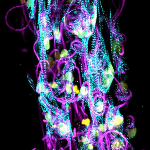
The team developed a microfabrication approach to produce “anisotropic myobundles”—highly aligned centimeter-long strips of stem cell-derived cardiac muscle tissue. They achieved this by blending naturally derived extracellular matrix (ECM) hydrogels with synthetic, cell-adhesive electrospun fiber segments.
To create a functional patch, they found that two critical components must work in tandem to guide the formation of functional myobundles:
“Maggie’s paper is centered on how to build engineered heart tissue at larger scales relevant to making a therapeutic patch,” Dr. Baker noted. “The study dives deeply into the nuanced aspects of how to seamlessly incorporate these structural fibers and determines exactly which supporting cell types are needed to drive functional tissue formation.”
The synergy between the lab-made fibers and the iCFs yielded exciting results. When combined, they formed highly aligned, three-dimensional tissue structures with enhanced cell spreading, superior myofibril formation, and synchronous contraction, providing a major step forward in biomanufacturing therapeutic heart tissue.
“This work identifies material and cell compositions that could be instrumental in developing a cardiac patch to restore heart function following a heart attack,” explained Jewett. “And as a result, helps advance the field toward a therapeutic approach to heal a currently terminal injury.”
With a robust, handleable patch in hand, the team is now gearing up for small animal studies towards eventual translational studies, bringing the medical community one step closer to repairing hearts without the need for heart transplants.