Innovative Biomaterial Scaffolds Enable Precise Recruitment and Study of Rare Antigen-Specific T Cells
A collaborative team led by U-M BME develops a controlled-release platform to advance immune cell research and unlock new possibilities in autoimmune disease monitoring.
For decades, researchers have been stymied by the challenge of studying the body’s rare, antigen-specific T cells—key players in immune defense and the progression of diseases such as multiple sclerosis (MS), cancer, and infections. Traditional approaches, such as blood draws and ex vivo expansion, often fail to collect sufficient numbers of these elusive cells or risk altering their function, thereby limiting scientific insight and therapeutic development.
Now, a multidisciplinary team of U-M biomedical engineers offers a new solution. In their latest publication, Aaron Morris, Assistant Professor, Biomedical Engineering, and fourth-year Ph.D. candidate Sydney Wheeler unveil a novel biomaterial platform engineered to recruit, enrich, and study antigen-specific T cells in vivo. This technology uses implantable polymer scaffolds that deliver disease-relevant antigens with unprecedented precision and control, enabling targeted collection of these rare immune cells directly from living tissue.
Built upon foundational work in immunological niche engineering, the Morris Lab’s new approach has the potential to revolutionize disease modeling, diagnostics, and researchers’ understanding of immune cell behavior in health and autoimmunity.
Studying the immune system’s response to disease is a cornerstone of biomedical engineering, yet one of its most valuable windows—rare antigen-specific T cells—has long been obscured. These T cells, crucial for the body’s defense and implicated in conditions ranging from infections to autoimmune disorders, are notoriously difficult to collect and analyze due to their exceptional scarcity in blood circulation.
“For years, researchers have relied on blood draws and expansion techniques to study antigen-specific T cells. These approaches are labor-intensive, costly, and at times, actually change the essential nature of the cells we’re trying to understand,” explained Dr. Morris, who leads the Morris Lab. “We wanted to rethink how we access and observe these critical cells in the context of disease.”
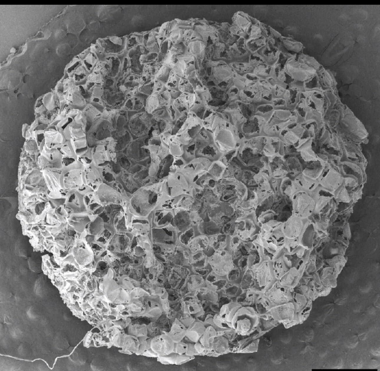
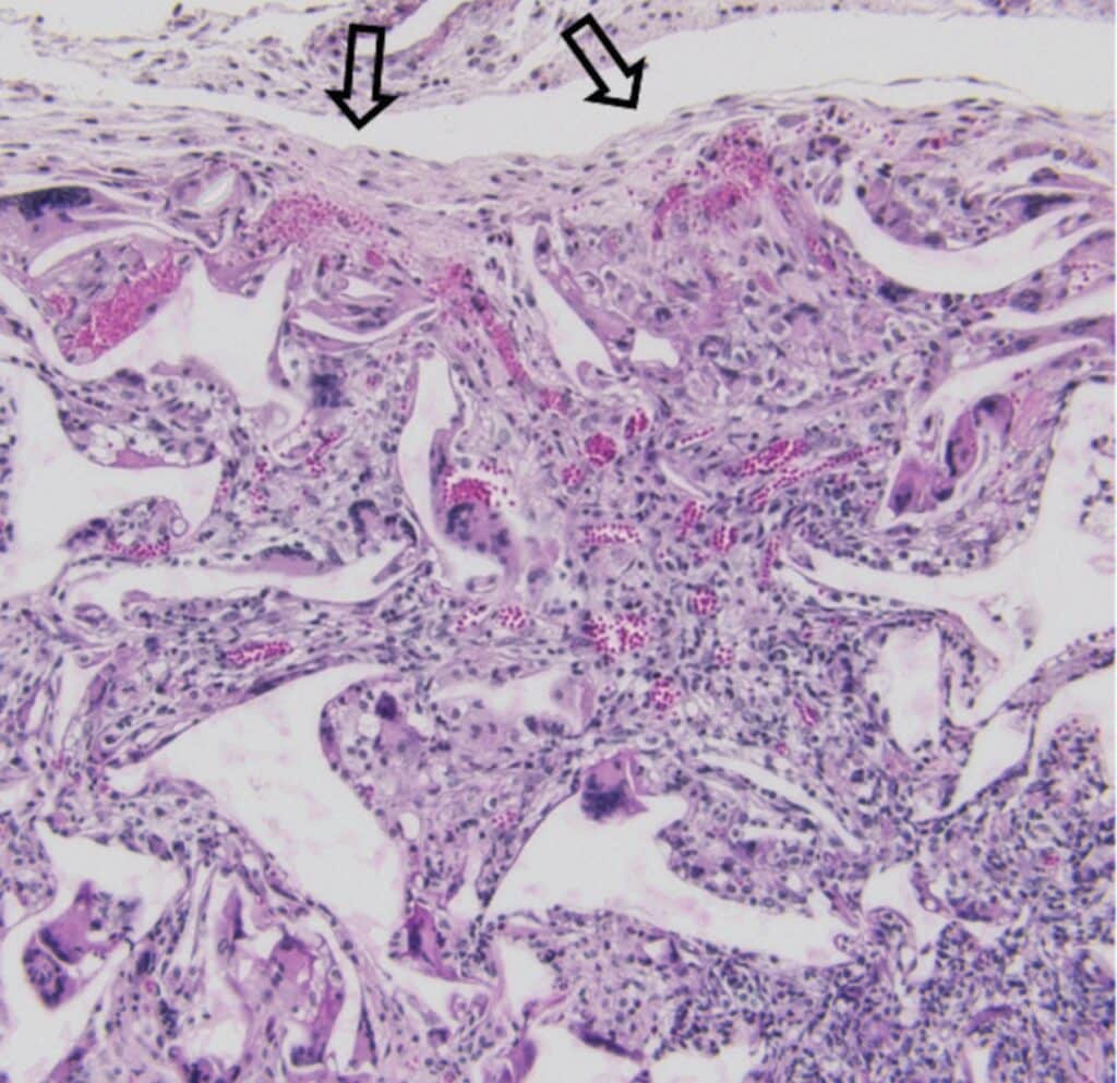
Enter the antigen-conjugated scaffold—a new class of implantable biomaterial designed by Morris, Ph.D. candidate Wheeler, and a collaborative cohort of graduate and undergraduate researchers. Their work, recently published in the Journal of Controlled Release, describes a technology that allows for precise, sustained delivery of disease-relevant antigens from a porous polymer scaffold implanted subcutaneously, which in turn draws in rare T cells at defined locations in vivo.
“It’s been a long time coming,” said Wheeler. “I’ve been working on this project since the early days of my Ph.D., and it builds directly on earlier work Professor Morris did during his postdoctoral research. The premise originally was to leverage implanted biomaterials to learn about the host immune system by recruiting immune cells to the site. But the challenge was always that the cells that were captured were predominantly innate immune cell populations—good for many studies, but not enough to reflect the full complexity of disease.”
Dr. Morris elaborated on the scientific roadblocks: “In our earlier studies, we saw exciting signs that immune cell patterns at the site of the scaffold reflected changes happening during disease, including models of multiple sclerosis. However, it was clear we weren’t capturing lots of antigen-specific T cells, which are central in autoimmunity. These cells exist as less than one in 100,000 in the blood, and harvesting and expanding them outside the body risks changing their biology. Our goal became finding a way to recruit these antigen-specific cells in vivo, in sufficient numbers and with minimal changes to their biology.”
A Precise and Modular Platform
Wheeler’s work represents a pivotal advance in scaffold chemistry. While previous approaches relied on simply entrapping antigens in scaffolds–often resulting in uncontrolled, rapid release–the U-M team used carbodiimide cross-linking to chemically bond peptide antigens directly to the backbone of PLG—a synthetic polymer already used in clinical devices. “By attaching the antigen to the polymer prior to making the scaffold, we can ensure precise loading and a slow, sustained release profile,” Wheeler explained. “This gives us the ability to finely tune the system for different diseases and targets, which is a huge advantage.”
Dr. Morris underscored the importance of control: “The amount of antigen delivered really matters biologically. We want to hit a sweet spot—enough to recruit the right T cells, but not so much as to cause rapid exhaustion or activation-induced cell death, which has affected other approaches. By weighing the polymer and knowing the efficiency of our chemical conjugation, we’re able to reproducibly manufacture scaffolds across experiments.”
Unlocking New Avenues for Immune Cell Research
Using animal models, the team showed that these antigen-conjugated scaffolds could successfully recruit rare CD4 T cells specific to antigens such as ovalbumin—a commonly used tool in immunology—and myelin protein fragments relevant to multiple sclerosis. The approach is highly modular; Wheeler noted, “We can target pretty much any disease where the relevant antigens are known, or use a mixture if there are multiple antigens. The system is tunable, and depending on what antigens you load, you can recruit different types of T-cell populations.”
Beyond multiple sclerosis, Dr. Morris sees broad implications. “You might want antigen-specific T cells for all kinds of reasons—studying infections, monitoring vaccine responses, designing cancer immunotherapies. Most tools right now require lots of blood and complicated ex vivo manipulations; here, we’re providing a minimally invasive way to enrich and study these key immune cells in vivo.”
Wheeler reflected on what makes the approach especially valuable in research: “T-cells are important for orchestrating many aspects of immunity, but the ones highly specific for a given antigen are extraordinarily rare in circulation. If we can create a local site to collect them—without expansion or alteration—we have a new opportunity to learn about disease mechanisms, develop therapeutics, and even monitor disease over time in animal models and potentially in patients.”
A Collaborative Effort with Broad Impact
Both Wheeler and Dr. Morris emphasized the collaborative nature of their research. “A number of undergraduate and graduate students contributed,” Wheeler said. “We also want to recognize Andrés Muñoz-Rojas from Rensselaer Polytechnic Institute (RPI), who has been a key collaborator and whose contributions are integral to this publication.”
Morris echoed this gratitude: “We have ongoing collaborations with Andrés Muñoz-Rojas at RPI, who contributed significantly to this work. Innovation in science happens best with collaboration, when folks with different backgrounds and strengths converge on a challenge.” The team also thanks Innovation Partnerships at U-M for their assistance with patent filing and technology transfer.
Looking Forward
While current results focus on technology development and animal models, the team has a clear vision for translation. The hope is that, with further work, antigen-conjugated scaffolds may provide clinicians and researchers with new tools for early detection of autoimmune, infectious, or cancerous diseases, and for tailoring therapies to individual patients.
“There’s potential here to use these tools for all kinds of immunological applications, including learning which antigens a person responds to,” Dr. Morris said. “We’ve filed a provisional patent, and we’re excited about both the basic science and the translational opportunities ahead.”
Wheeler summarized the spirit of the project: “It’s a platform—a building block. We wanted to see if we could develop a system where we deliver antigens and actually see the specific cells we care about. We’ve validated that, and from here, the directions are wide open.”