New Capsule Technology Shows Promise for Restoring Lost Ovarian Function due to Cancer Treatments
U-M Biomedical Engineering researchers make strides toward improving quality of life for pediatric cancer survivors.
5–8 minutes
U-M Biomedical Engineering researchers make strides toward improving quality of life for pediatric cancer survivors.
For thousands of young cancer survivors, life after remission can bring a new set of challenges—especially for girls and teens whose treatments damage the ovaries. Standard therapies such as chemotherapy and radiation, while life-saving, often harm thea non-renewable reserve of ovarian follicles. These tiny structures play a key role in producing the hormones needed for puberty and health. Without them, survivors face premature ovarian insufficiency (POI), leading to not just infertility, but also a host of issues—ranging from bone fragility and heart problems to neurological and immune system complications.
Until now, the only way to treat POI in adolescents has been hormone replacement therapy (HRT), which supplies only two of the ovarian hormones, estradiol and progesterone. But there’s a major catch: HRT can’t replicate the natural ebb and flow of hormones between a healthy ovary and the rest of the body. Instead, it delivers fixed amounts of hormones, missing the complex, finely tuned feedback system that real ovaries provide. This “one-size-fits-all” approach significantly increases the risks for musculoskeletal, cardiovascular, neurological and metabolic disease, while the long-term safety remains unclear.
Taking Inspiration from Nature
“Currently, the only option treating these patients is an off-label prescription of HRTn, which is not designed for the initiation of puberty,” explained Ariella Shikanov, Professor, Biomedical Engineering and Obstetrics and Gynecology.
Margaret Brunette, Ph.D., a former graduate student in the Shikanov lab has pioneered a new approach for encapsulation of human ovarian tissue in a hydrogel-based immune-isolating capsule that may allow donor ovarian tissue to be transplanted, without fear of immune rejection or the need for lifelong immunosuppression.
“We investigated if we could take a small piece of ovarian tissue, implant it in an organism or animal model that doesn’t have functioning ovaries, and see if this tissue can function as a healthy host ovary,” Dr. Shikanov said.
Historically, autologous transplants of ovarian tissue—often from the person themselves—have helped restore fertility and normal hormone production. But for pediatric cancer survivors, this carries risks. Cryopreserving ovarian tissue before cancer therapy isn’t common practice, and for some children (especially those with blood cancers), using their own tissue could reintroduce harmful cancer cells.
Using donor tissue would be safer, but requires suppressing the immune system to avoid rejection—hardly ideal for growing kids. So, the U-M team’s innovative workaround is in the form of a hydrogel-based capsule that acts as an immune shield. This flexible, soft jello-like “bubble” allows nutrients and hormones to flow in and out, but prevents infiltration of immune cells, protecting the transplanted ovarian tissue from rejection.
“What this paper is showing is that encapsulation and isolation of the tissue from blood supply and the immune system does not negatively affect its function,” said Dr. Shikanov. In other words, even though the tissue is not connected to the animal’s blood supply, it still thrives and produces hormones within the capsule similar to the non-encapsulated control tissue. Estradiol levels—a key marker for functioning ovaries—reached healthy physiological amounts. “We could restore ovarian endocrine function and reach physiological levels of circulating hormones, specifically estradiol, and it was as good as non-encapsulated tissues, confirming that the idea of encapsulation works.”
Proof of Concept—Letting Ovarian Tissue Thrive
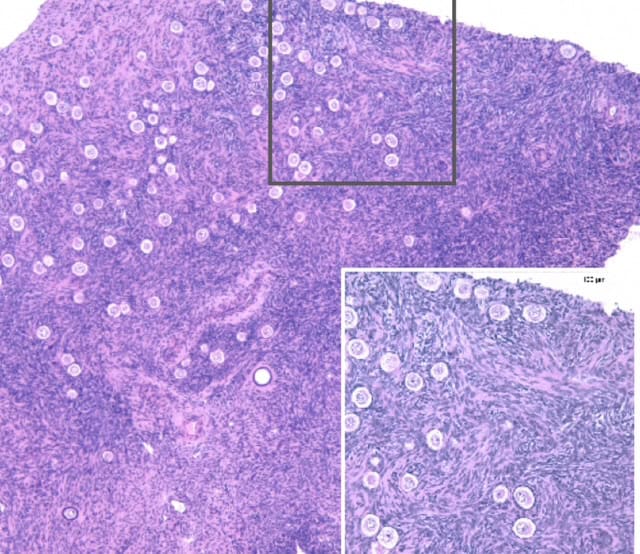
In their latest study, the U-M team tested this approach in immunodeficient mice that had their ovaries removed. They encapsulated and implanted human ovarian tissue from deceased donors over several months; they monitored the mice using hormone measurements, daily checks for estrous cycles (the mouse equivalent of menstrual cycles), and analyzed retrieved grafts.
The results were remarkable: within 12 weeks, the mice began having regular estrous cycles—indicating that the implanted tissue was communicating with the brain hormone control centers, just like a natural ovary. Large follicles developed inside the capsule, producing increasing levels of estradiol (a key estrogen hormone) that reached healthy levels. In other words, the donor tissue not only survived but restored hormone function—matching non-encapsulated controls. This process echoed the natural biology of human ovarian follicles, which need several months to mature and require supporting matrices.
Importantly, these hormone cycles weren’t artificially induced but emerged naturally, showing the tissue had integrated with the mouse’s own endocrine system. The study also confirmed the presence of other vital cell types needed for hormone production—meaning the whole ovarian “machinery” was running.
Looking Forward—A Safer Path to Restoring Female Health
These results mark a significant step toward a safer way to help young cancer survivors regain their full spectrum of health. While the study was conducted in immunocompromised mice (meaning capsules didn’t have to fend off active immune systems), previous research with animal tissue suggests the capsule can protect against immune rejection. Next steps will include testing in immune-competent models, assessing long-term outcomes, and exploring ways to fine-tune the process for real-world treatment.
Though challenges remain—such as ensuring consistent follicle counts and full hormone profiles—the U-M team’s hydrogel capsule brings hope. One day, it could mean that “beating cancer” no longer comes at the cost of losing natural hormone function. By combining ingenuity and compassion, biomedical engineers are moving closer to restoring not just health, but also the future possibilities for the kids and families who need it most.
Next Steps
The research, now published in Science Advances, is in the process of moving to the next stage: non-human primate studies, thanks to another R01 grant from the NIH. “We travelled to the Oregon Non-Human Primate Center, and started our first large non-human primate studies, where we grafted allogeneic ovarian tissue into healthy, non-immune compromised monkeys,” said Dr. Shikanov. “Right now, we are running a large non-human primate study. I want to acknowledge the Frankel Initiative and the NIH, for their support of our preclinical studies. We plan to submit an application to the FDA with a goal of receiving approval to start first-in-human clinical trials.”
Students in the Shikanov lab are currently working to make the capsule even better—optimizing nutrient flow, expanding to other species, and testing whether they can cryopreserve the capsule for easier shipping,longer shelf life and accessibility.
Hope on the Horizon
Dr. Shikanov is clear on her vision: “The end goal is to bring these advances to the people in the clinic.” For young cancer survivors and their families, this means hope for a future where “beating cancer” no longer means losing out on the chance for natural puberty and lifelong hormonal health.
Acknowledgments:
This work was supported by a number of generous grants and contributions, including the National Institutes of Health (R01HD104173, R01EB022033), Eunice Kennedy Shriver NICHD/NIH (R24HD102061, Z1A HD009005), T32DE007057 and T32HD079342 training grants, and the Musculoskeletal Transplantation Foundation (MTF) Biologics Research Grant. Hormone analysis was performed by the University of Virginia Center for Research in Reproduction Ligand Assay and Analysis Core. Sample processing for tissue sectioning was handled by the University of Michigan Dental Histology Core, with whole slide scanning provided by the University of Michigan ULAM Pathology Core. The Frankel Innovation Initiative, ARPA-H, and numerous student contributors in the Shikanov lab also played invaluable roles in making this research possible.
For more news and updates:
Follow us on LinkedIn, and our funding sources Eunice Kennedy Shriver National Institute of Child Health and Human Development (NICHD), National Institute of Biomedical Imaging and Bioengineering (NIBIB), MTF biologics and the Frankel Innovation Initiative.