A ‘Communication Breakdown’ During General Anesthesia
What happens to the brain during general anesthesia?

What happens to the brain during general anesthesia?
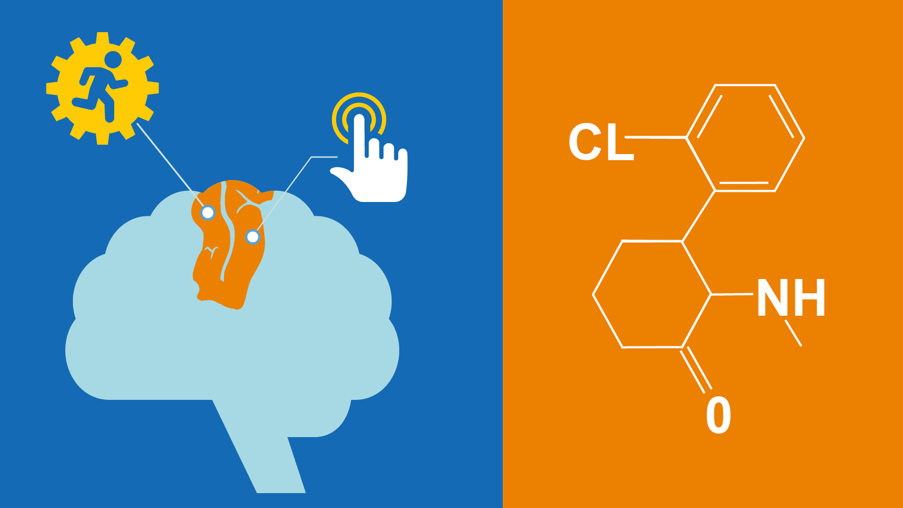
When ketamine is used for general anesthesia, two connected parts of the cortex turn to “isolated cognitive islands.”
It’s a topic that has long captivated doctors, scientists and the public — what exactly happens in your brain when you’re oblivious on the operating table?
Some anesthesia drugs work in a straightforward manner by dampening down neurons in the brain. The mechanism of one anesthetic, however, has proved elusive: ketamine.
Certain doses of ketamine induce general anesthesia, though brain activity can still be robust, says Cynthia Chestek, Ph.D., co-senior author of a new study in NeuroImage.
Ketamine is used often in patient care and in laboratory settings. The new paper examines the neurological mechanisms at work during ketamine anesthesia.
Co-senior authors Chestek and anesthesiologist George Mashour, M.D., Ph.D., led the research team, which took precise measurements down to the level of neurons in animal models.
“We found that general anesthesia reflects a communication breakdown in the cortex, even though sensory information is getting processed,” Mashour says. “But the processing appears to occur in isolated cognitive islands.”
Turns out, two adjacent parts of the brain that work together in the waking state simply stop talking to each other under general anesthesia. When awake, communication between the primary somatosensory cortex and the primary motor cortex is critical to normal function.
“This supports the idea that what anesthesia does to cause unconsciousness is interrupt communication between brain areas, stopping the processing of higher-level information,” says first author Karen Schroeder, a doctoral candidate in the U-M Department of Biomedical Engineering. “This was the first time anyone directly observed the interruption between the two areas using individual neurons.”
Chestek’s biomedical engineering lab focuses on brain machine interfaces, recording activity of neurons and reading motor commands and sensory information in real time.
“This turned out to be really useful for the researchers in anesthesiology,” Chestek says. So her team got on board to measure both areas of the brain, which kept firing during anesthesia.
“As soon as we injected ketamine, the sensory information disappeared from the motor cortex. Normally these areas are tightly connected.”
The group plans to continue this work, turning next to investigate the level of anesthesia at which these changes in communication start to occur. They’re also looking into what the groups of neurons are doing under anesthesia when they are still active but no longer communicating with each other.
“These insights could potentially improve our ability to monitor patients’ level of consciousness,” Schroeder says.
Funding for the work came from the National Institutes of Health.